THE USE OF THE PAPANICOLAOU STAIN TO ROUTINE HISTOLOGICAL EXAMINATION: TEACHING THE BASIC MICROSCOPY OF SQUAMOUS CELL CARCINOMA
DOI:
https://doi.org/10.34019/2177-3459.2017.v9.24041Keywords:
Papanicolaou. Staining. Histopathology. Squamous cell carcinoma.Abstract
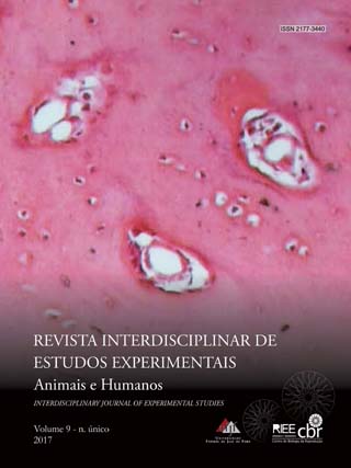
Introduction: The understanding of microscopy classes is imperative to the student in human pathology. Objective: To prepare the student to observe and understand, through tinctorial recognition, the main histologic features of Papanicolaou-stained well-differentiated carcinomas. Methods: A selected case of a well-differentiated carcinoma of the tongue was used for a Papanicolaou stain. Results: The Papanicolaou-stained slides showed, basically, basal cells in a faint to a dark blue color. The stratum spinosum stained light pink to a faint blue-green and the keratin layer and the keratin pearls stained bright orange. The connective tissue stained light green. Conclusion: The use of Papanicolaou stain was found very convenient as the beginner in histopathology can perform a ready identification of the well-differentiated squamous cell carcinoma aspects.
Downloads
Downloads
Published
Issue
Section
License
Autores que publicam nesta revista concordam com os seguintes termos:- Autores mantém os direitos autorais e concedem à revista o direito de primeira publicação, com o trabalho simultaneamente licenciado sob a Creative Commons Attribution License que permitindo o compartilhamento do trabalho com reconhecimento da autoria do trabalho e publicação inicial nesta revista.
- Autores têm permissão e são estimulados a citar e distribuir seu trabalho (ex.: em repositórios institucionais, página pessoal, trabalhos científicos, etc) desde que citada a fonte (referência), já que isso pode gerar produtividade para os autores, bem como aumentar o impacto e a citação do trabalho publicado.
