
Fractal dimension and lacunarity as bone quality parameters in the mandible of healthy individuals: a cone-beam computed tomography analysis
DOI:
https://doi.org/10.34019/1982-8047.2021.v47.32583Keywords:
Fractals, Mandible, Cone-Beam Computed TomographAbstract
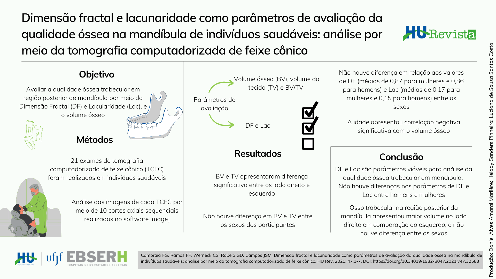
Introduction: Bone quality can be assessed trough a texture analysis in imaging exams by Fractal dimension (FD) and lacunarity (Lac) measurements. DF represents the complexity of a fractal and Lac means distribution and size of the empty spaces. Objective: To evaluate trabecular bone quality through FD, Lac, and bone volume in the posterior region of the mandible using cone-beam computed tomography (CBCT) in healthy individuals, also comparing the results between males and females. Materials and Methods: 21 CBCT exams were used. The software ImageJ was used with ten sequential axial slices were selected for each patient, and the region of interest (ROI) was defined between the lower canine and first molar in both sides. The parameters were evaluated: a) Bone Volume (BV), Tissue Volume (TV) and BV/TV by binarization process; b) FD and Lac by the box-counting method in greyscale, without binarization. Results: When comparing the sides, BV and TV had a significant difference (p=0.02 e 0.01, respectively) with higher values on the right side. When comparing the sexes, there was no significant difference. The values for FD had an average of 0.87 for females and 0.86 for males. Lac values were 0.17 for females and 0.15 for males, in average. The age had negative significant correlation with bone volume. Conclusion: DF e Lac are viable parameters for analyse to trabecular bone quality in the mandible. Values were found lower than 1 for FD and lower than 0.17 for Lac in healthy individuals. FD and Lac had no difference between males and females. The trabecular bone in the posterior region of the mandible had higher values on the right side when compared to the left side, and there was no difference between sexes.
Downloads
References
Farlay D, Boivin G. Bone mineral quality. Osteoporosis. 2012; 953-78.
Chavassieux P, Seeman E, Delmas PD. Insights into material and structural basis of bone fragility from diseases associated with fractures: how determinants of the biomechanical properties of bone are compromised by disease. Endocr Rev. 2007; 28(2):151-64.
Irie MS, Rabelo GD, Spin-Neto R, Dechichi P, Borges JS, Soares PBF. Use of micro-computed tomography for bone evaluation in dentistry. Braz Dent J. 2018; 29(3):227-38.
Geraets WG, Van Der Stelt PF. Fractal properties of bone. Dentomaxillofac Radiol. 2000; 29(3):144-53.
Yasar F, Akgunlu F. Evaluating mandibular cortical index quantitatively. Eur J Dent. 2008; 2(4):283-90.
Alman AC, Johnson LR, Calverley DC, Grunwald GK, Lezotte DC, Hokanson JE et al. Diagnostic capabilities of fractal dimension and mandibular cortical width to identify men and women with decreased bone mineral density. Osteoporos Int. 2012; 23(5):1631-6.
Kavitha MS, Park SY, Heo MS, Chien SII. Distributional variations in the quantitative cortical and trabecular bone radiographic measurements of mandible, between male and female populations of Korea, and its utilization. PLoS One. 2016; 11(12):e0167992.
Rabelo GD, Camillo-Coutinho C, Kowalski LP, Portero-Muzy N, Roux JP, Chavassieux P et al. Evaluation of cortical mandibular bone in patients with oral squamous cell carcinoma. Clin Oral Investig. 2018; 22(2):783-90.
Melo RHC, Conci A. How Succolarity could be used as another fractal measure in image analysis. Telecommun Syst. 2013; 52(3):1643-55.
Gumussoy I, Miloglu O, Cankaya E, Bayrakdar IS. Fractal properties of the trabecular pattern of the mandible in chronic renal failure. Dentomaxillofac Radiol. 2016; 45(5):20150389.
Rabelo GD, Roux JP, Portero-Muzy N, Gineyts E, Chapurlat R, Chavassieux P. Cortical fractal analysis and collagen crosslinks content in femoral neck after osteoporotic fracture in postmenopausal women: comparison with osteoarthritis. Calcif Tissue Int. 2018; 102(6):644-50.
Rodrigues GHC, Rodrigues VA, Barros SM, Romeiro RL, Souza DM. Computed tomography X panoramic radiography in the evaluation pre-surgical in implantology. Innov Implant J, Biomater Esthet. 2012/2013; 7(8):126-31.
Torres SR, Chen CSK, Leroux BG, Lee PP, Hollender LG, Schubert MM. Fractal dimension evaluation of cone beam computed tomography in patients with bisphosphonate-associated osteonecrosis. Dentomaxillofac Radiol. 2011; 40(8):501-5.
Kato CNAO, Barra SG, Tavares NPK, Amaral TMP, Brasileiro CB, Mesquita RA et al. Use of fractal analysis in dental images: a systematic review. Dentomaxillofac Radiol. 2020; 49(2):20180457.
Monteiro LH, Conci A. Reconhecimento de placas de veículos utilizando processamento de imagem. Engevista. 2003; 5(10):31-43.
Shrout MK, Potter BJ, Hildebolt CF. The effect of image variations on fractal dimension calculations. Oral Surg Oral Med Oral Pathol Oral Radiol Endod. 1997; 84(1):96-100.
Suomalainen A, Pakbaznejad Esmaeili E, Robinson S. Dentomaxillofacial imaging with panoramic views and cone beam CT. Insights Imaging. 2015; 6(1):1-16.
Özalp Ö, Tezerişener HA, Kocabalkan B, Büyükkaplan UŞ, Özarslan MM, Şimşek Kaya G, Altay MA, Sindel A. Comparing the precision of panoramic radiography and cone-beam computed tomography in avoiding anatomical structures critical to dental implant surgery: a retrospective study. Imaging Sci Dent. 2018; 48(4):269-75.
Helkimo E, Carlsson GE, Helkimo M. Chewing efficiency and state of the dentition. A methodologic study. Acta Odontol Scand. 1978; 36(1):33-41.
Oettlé AC, Becker PJ, de Villiers E, Steyn M. The influence of age, sex, population group, and dentition on the mandibular angle as measured on a South African sample. Am J Phys Anthropol. 2009; 139:505-11.
White TD, Black MT, Folkens PA. Human osteology. 3rd. ed. Amsterdam: Elsevier, Academic Press; 2011.
Rehman MT, Hoyland JA, Denton J, Freemont AJ. Age related histomorphometric changes in bone in normal British men and women. J Clin Pathol. 1994; 47(6):529-34.
Giodano V, Franco JS, Koch HA, Labronici PJ, Pires RES, Amaral NP. Age-related changes in bone architecture. Rev Col Bras Cir. 2016; 43(4):276-85.
Downloads
Published
How to Cite
Issue
Section
License
Copyright (c) 2021 Gabrielle Cambraia Faria, Fernanda Ramos de Faria, Carolina de Sá Werneck, Gustavo Davi Rabelo, Marcio José da Silva Campos

This work is licensed under a Creative Commons Attribution 4.0 International License.
Cessão de Primeira Publicação à HU Revista
Os autores mantém todos os direitos autorais sobre a publicação, sem restrições, e concedem à HU Revista o direito de primeira publicação, com o trabalho licenciado sob a Licença Creative Commons Attribution que permite o compartilhamento irrestrito do trabalho, com reconhecimento da autoria e crédito pela citação de publicação inicial nesta revista, referenciando inclusive seu DOI.